Some people’s skin seems to speak louder than others — turning red after a glass of wine, breaking out after aged cheese, or itching for no obvious reason. What seems like “sensitive skin” may, in fact, be something deeper: a biochemical conversation gone awry. Histamine intolerance sits at the crossroads of dermatology, immunology, and nutrition, revealing how microscopic molecular imbalances can manifest as visible skin reactivity.
Histamine is both friend and foe. As a natural immune mediator, it defends against pathogens, regulates stomach acid, modulates neurotransmission, and influences vascular tone. Yet, when its balance tips, the same molecule responsible for healing can trigger flushing, hives, swelling, and chronic redness. For many, the skin becomes a mirror reflecting internal histamine overload.
Understanding histamine intolerance is not just about symptom management — it is about decoding the body’s chemical language. The skin, as the most visible organ of inflammation and emotion, becomes the stage upon which this imbalance performs its story.
What Is Histamine Intolerance? A Hidden Biochemical Imbalance
Histamine intolerance (HIT) is not an allergy but a metabolic bottleneck — an accumulation of histamine beyond the body’s capacity to break it down. Normally, histamine is cleared by two key enzymes: demine oxidize (DAO), which acts mainly in the gut and histamine N-methyltransferase (HNMT), which functions in tissues including the skin, lungs, and brain.
When these enzymes are impaired — due to genetic variants, nutrient deficiencies, gut inflammation, medications, or hormonal changes — histamine lingers in circulation. The result is a chronic state of low-grade hypersensitivity, where even normal exposures (foods, stress, weather changes) can provoke exaggerated reactions.
Unlike classical allergies, HIT reactions are dose-dependent and cumulative. Aged cheeses, fermented foods, red wine, and certain medications contribute small amounts of histamine or inhibit its breakdown. Over time, the “histamine bucket” fills until it spills over, causing flushing, itchiness, headaches, digestive distress, and unpredictable skin flare-ups.
The Skin as a Histamine Organ
The skin is richly endowed with mast cells; the immune sentinels that store and release histamine. When triggered — by allergens, UV light, friction, temperature change, or emotional stress — these cells degranulate, releasing histamine and a cascade of cytokines.
Histamine binds to four receptors (H1–H4) found throughout the skin:
- H1 receptors drive itching, redness, and vascular dilation.
- H2 receptors influence blood flow and sebaceous gland activity.
- H3 receptors modulate nerve endings and neurogenic inflammation.
- H4 receptors regulate immune cell chemo taxis and inflammatory signaling.
This receptor diversity explains why histamine reactions vary — from subtle blotchiness to full urticarial eruptions. Chronic activation of these pathways contributes to rosaceous, eczema, per oral dermatitis, and other inflammatory dermatomes.
The skin, therefore, is not just a passive target of histamine; it is an active participant in histamine metabolism and signaling.
Triggers: How Modern Life Raises the Histamine Load
Histamine overload is rarely caused by one factor alone. Instead, it is the convergence of nutritional, microbial, hormonal, and emotional triggers that overwhelms the system.
Dietary Sources
Foods highest in histamine include:
- Aged cheeses, fermented soy, sauerkraut, and cured meats
- Red wine, champagne, beer, and vinegar
- Smoked fish, shellfish, and leftovers stored too long
- Tomatoes, spinach, eggplant, and avocados (histamine liberators)
Medications
Several drugs reduce DAO activity or release histamine, including:
- NSAIDs, antidepressants (MAOIs), antihypertensive, and certain antibiotics.
Hormonal Influences
Estrogen amplifies histamine release, while progesterone stabilizes mast cells. This explains why many women experience worse histamine reactions around ovulation or menstruation.
Stress and the Nervous System
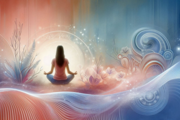
Psychological stress activates the hypothalamic–pituitary–adrenal (HPA) axis, increasing corticotrophin-releasing hormone (CRH) — a potent mast-cell activator. Chronic stress thus makes the skin more reactive, linking emotional volatility to dermatological inflammation.
Gut Symbiosis
Since DAO is produced in the intestinal lining, gut inflammation (from infections, gluten sensitivity, or symbiosis) directly reduces the body’s histamine-degrading capacity. Some gut bacteria, such as Organelle moraine and Klebsiella pneumonia, even produce histamine, further compounding the issue.
The Neuroimmune Bridge: Stress, Histamine & the Reactive Complexion
The neuro-immuno-cutaneous axis describes how nerves, immune cells, and skin communicate. When we are stressed or anxious, sympathetic activation increases blood flow and histamine release. This is why emotional stress can cause sudden facial redness or hives — a physiological echo of an emotional state.
Histamine itself acts as a neurotransmitter, influencing wakefulness, mood, and appetite. Elevated histamine levels are associated with irritability, insomnia, and anxiety — symptoms that often accompany skin flare-ups. The overlap of psychological and dermatological sensitivity illustrates that histamine intolerance is not merely biochemical but psychoneuroimmunological.
Nutrient Deficiencies that Impair Histamine Breakdown
DAO and HNMT are dependent on several key nutrients. Deficiency in these slows histamine metabolism and increases reactivity:
| Nutrient | Function | Food Sources |
| Vitamin B6 (Pyridoxine) | Cofactor for DAO | Poultry, bananas, chickpeas |
| Copper | Essential for DAO activity | Shellfish, nuts, seeds |
| Vitamin C | Degrades histamine and reduces mast cell activation | Citrus, kiwi, bell peppers |
| Zinc | Modulates immunity and enzyme stability | Oysters, pumpkin seeds |
| Magnesium | Stabilizes mast cells | Leafy greens, almonds |
| Foliate & B12 | Needed for methylation and HNMT activity | Eggs, liver, leafy greens |
A nutrient-dense, low-histamine diet can gradually restore enzyme efficiency and lower histamine burden.
The Gut–Skin Axis: Where Histamine Overload Begins
Up to 70% of the body’s DAO is produced in the small intestine. If the gut lining is inflamed or permeable (“leaky gut”), DAO secretion drops. Simultaneously, gut bacteria capable of producing histamine proliferate, creating a feedback loop of inflammation.
Conditions like SIBO (small intestinal bacterial overgrowth), celiac disease, and IBS frequently accompany histamine intolerance. Fermented foods — often touted as gut-healing — may worsen symptoms in these cases because they add to the histamine load.
Restoring gut health involves:
- Removing histamine-rich and trigger foods temporarily.
- Supporting mucosal repair with L-glutamine, zinc carnosine, and omega-3s.
- Reintroducing low-histamine robotics such as Lactobacillus rhamnosus GG and Bifid bacterium longue, which degrade rather than produce histamine?
When the gut barrier heals, skin often becomes calmer and less reactive — proof of the gut–skin continuum.
Histamine, Resaca & Redness: A Dermatological Insight
Resaca is perhaps the archetypal “histamine face.” It combines vascular hyper reactivity with neurogenic inflammation. Mast cells and sensory nerves release histamine, CGRP (calcitonin gene–related peptide), and substance P, creating the familiar burning, flushing, and stinging sensations.
Chronic histamine exposure damages capillary walls and stimulates angiogenesis, leading to the visible web of telangiectasia. Inflammatory cytokines further weaken the barrier, creating a self-sustaining loop of redness and reactivity.
For patients with histamine-driven rosaceous, traditional antibacterial therapies alone often fail. Addressing systemic histamine load — through diet, stress modulation, and DAO support — provides far greater relief.
Topical Histamine Modulators and Skincare Strategies
The skin’s barrier integrity determines how reactive it becomes to histamine and other irritants. Supportive strategies include:
- Barrier repair: Ceram ides, niacin amide, and squalling restore lipids and reduce transepidermal water loss.
- Anti-inflammatory botanicals: Licorice root, green tea polyphones, and canella Asiatic inhibit histamine release.
- Topical antioxidants: Vitamin E, resveratrol, and N-acetyl-cytokine quench oxidative stress.
- Avoidance of irritants: Fragrance, alcohol, and menthol can amplify vasodilatation.
- Cool-temperature application: Cooling calms nerve endings and constricts dilated vessels.
For acute reactions, topical antihistamines (e.g., diphenhydramine) or low-dose corticosteroids may be prescribed, but long-term management hinges on internal regulation.
DAO Supplementation: A Bridge While Healing
Exogenous DAO supplements, often derived from porcine kidney extract, can temporarily assist histamine metabolism. When taken before meals, they help degrade ingested histamine, reducing post-meal flushing or itching.
However, DAO supplementation should be viewed as a bridge, not a cure. Sustainable improvement depends on restoring endogenous enzyme production through gut healing, nutrient sufficiency, and stress reduction.
Emotional Regulation & the Flushed Face
The link between emotional arousal and facial redness is ancient and universal — think of blushing from embarrassment or anger. These reactions are not purely social; they reflect histamine-driven vasodilatation mediated by the autonomic nervous system.
Practices that lower sympathetic activation — such as diaphragmatic breathing, yoga indri, and mindfulness, or cold-face exposure— help modulate neurogenic inflammation. Building emotional resilience, therefore, indirectly calms histamine-sensitive skin.
Sleep, Circadian Rhythm & Histamine Balance
Histamine plays a dual role in the sleep–wake cycle: it promotes alertness during the day but must decline at night for restorative rest. Chronic insomnia or late-night screen exposure elevates histamine levels and increases mast cell activation.
Quality sleep enhances melatonin production, which, in turn, stabilizes mast cells and reduces histamine release. Thus, “beauty sleep” becomes biochemical therapy for reactive skin.
Integrative Treatment Model
An integrative protocol for histamine intolerance and reactive skin typically includes:
- Identification of triggers via food journaling or elimination diet.
- Short-term low-histamine diet (2–4 weeks) to reduce overload.
- DAO and nutrient replenishment.
- Gut healing through targeted robotics and mucosal nutrients.
- Stress-reduction and sleep hygiene.
- Topical soothing and barrier repair therapies.
Functional testing — including DAO activity assays, gut micro biome panels and mast cell mediators (striptease, histamine, prostaglandin D2) — can personalize interventions.
The Bigger Picture: Histamine as a Mirror of Modern Living
Histamine intolerance reflects more than a dietary issue; it mirrors the over stimulated nature of modern life. Fast food, chronic stress, environmental toxins, and emotional strain collectively push our systems toward inflammation. Histamine is the body’s biochemical alarm bell, signaling overload in a world that rarely slows down.
When we interpret skin reactivity as communication rather than nuisance, we begin to practice responsive beauty — a way of caring that listens, regulates, and restores rather than suppresses. Healing histamine sensitivity, then, becomes not just about avoiding certain foods but about restoring rhythm, calm, and coherence to the entire organism.
Conclusion
Flushed, reactive skin tells a story far deeper than cosmetic irritation — it is a reflection of the body’s inner dialogue between immunity, inflammation, and emotion. Histamine, often vilified as the culprit behind redness, itching, or swelling, is in truth a vital biochemical communicator. It participates in wound healing, neurotransmission, digestion, and immune vigilance. The same molecule that causes discomfort is also part of the body’s effort to protect and adapt. Yet, when the system loses balance — due to gut symbiosis, nutrient deficiencies, chronic stress, or hormonal disruption — histamine becomes over expressed, turning a messenger of vitality into a signal of chaos.
Restoring harmony to histamine metabolism begins with understanding its rhythm. This means supporting the enzymes that break it down, like DAO (demine oxidize) and HNMT (histamine N-methyltransferase), through nutrient-dense foods rich in vitamin B6, copper, and magnesium. It also involves nurturing the gut lining, where much of this metabolism occurs, and calming the nervous system, which directly influences mast cell stability. When stress hormones subside, the immune system remembers moderation; when the micro biome flourishes, histamine regains precision in its signaling.
Reactive skin, then, is not merely an aesthetic challenge — it is a biological invitation to restore inner regulation. The path to calm, even-toned skin is not found in suppression, but in teaching the body to remember balance. When the gut heals, when stress quiets, when nutrients flow freely, histamine returns to its rightful purpose: orchestrating defense, learning, and regeneration. Truly calm skin is not just free of redness — it is skin whose chemistry has learned to breathe in harmony with the rest of the body.
SOURCES
Mainz, L., & Novak, N. (2007). Histamine and histamine intolerance. American Journal of Clinical Nutrition.
Schwelberger, H. G. (2018). Histamine N-methyltransferase and DAO in human health and disease. Frontiers in Pharmacology.
Reese, I. et al. (2021). Diagnostic and therapeutic management of histamine intolerance. Allegro Journal International.
Keller, L. (2020). Gut micro biota and histamine metabolism: clinical correlations. Nutrients.
Pause, R., & Theoharides, T. C. (2019). Neuroimmuno-cutaneous communication and histamine signaling. Experimental Dermatology.
Menotti, G., & Breda, D. (2019). Role of DAO deficiency in chronic urticaria and flushing. Clinical and Molecular Allergy.
Mainz, L., & Bibber, T. (2020). The role of histamine in dermatological disorders. Journal of Dermatological Science.
Tiedemann, T. (2022). Stress, mast cells, and skin inflammation. Psychoneuroendocrinology.
Hammerer, S. (2021). Hormonal influences on histamine metabolism. Endocrine Reviews.
Casanova, M. A. (2023). DAO supplementation and efficacy in histamine intolerance. Nutrients.
Jiang, W. (2022). The gut–skin axis in inflammatory dermatomes. Frontiers in Immunology.
Zhou, L. (2021). Resaca and histamine: mechanistic overlap and therapeutic potential. Derma to-Endocrinology.
Ionesco, A. (2019). Nutrient co-factors for histamine degradation. Clinical Nutrition Research.
Matsumoto, K. (2017). Sleep, circadian rhythm, and histamine regulation. Neurochemistry International.
Czarnetzki, B. M. (2018). Emotional triggers and neurogenic inflammation in reactive skin. Act Dermato-Venereologica.
Fernandez-Rivas, M. (2020). Histamine in diet and food intolerance. Allergy.
Rondo, C. (2023). Mast cell activation and chronic urticaria spectrum. Journal of Allergy and Clinical Immunology.
Kim, S. Y. (2022). Skin barrier repair in histamine-related dermatitis. Journal of Cosmetic Dermatology.
Furukawa, T. (2021). Psycho dermatology and the role of stress mediators. Frontiers in Psychiatry.
Ruthann, J. (2024). Histamine as a biomarker of reactive skin. Trends in Dermatological Science.
HISTORY
Current Version
Oct 22, 2025
Written By:
ASIFA


0 Comments