Introduction
Obesity is traditionally viewed as a homogeneous condition characterized by excess adiposity, systemic metabolic disruption, and a heightened risk for chronic diseases. However, advances in metabolic medicine, adipose-tissue biology, immunology, and endocrine research have revealed that obesity is not a single uniform phenotype. Instead, at least two major biological subtypes are now recognized:
These categories reflect distinct differences in inflammatory tone, adipose-tissue function, immune cell activation, hormonal responses, metabolic flexibility, and cardio metabolic risk.
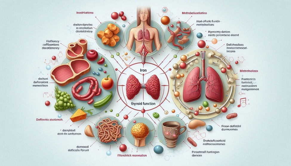
The Inflammatory Obesity Phenotype (IOP) is characterized by visceral adiposity, adipose-tissue macrophage infiltration, elevated pro-inflammatory cytokines, impaired insulin signaling, dysfunctional adipokine secretion, mitochondrial stress, and increased cardio metabolic risk. Individuals with IOP tend to exhibit higher levels of IL-6, TNF-α, MCP-1, CRP, and altered levels of lepton, adiponectin, and resisting, creating a metabolic environment that accelerates insulin resistance, hepatic statuses, and endothelial dysfunction.
Conversely, Metabolically Healthy Obesity (MHO) describes individuals with excess body fat but without the metabolic abnormalities typically associated with obesity, such as dyslipidemia, hypertension, chronic inflammation, or insulin resistance. These individuals often maintain better adipose-tissue expandability, higher adiponectin levels, lower visceral fat accumulation, preserved mitochondrial function, and favorable inflammatory markers.
However, MHO is not universally stable; many individuals transition from MHO to IOP over time, particularly under the influence of chronic stress, sleep disturbances, aging, nutrient imbalance, or environmental triggers. Understanding the mechanistic differences between these phenotypes is essential for designing precision nutrition strategies, anti-inflammatory dietary patterns, chronobiological eating schedules, and targeted lifestyle interventions.
This article explores—at a deep, mechanistic level—the biological divergence between Inflammatory Obesity and Metabolically Healthy Obesity, highlighting:
- Adipose-tissue immunology
- Inflammatory signaling
- Adipokine patterns
- Gut micro biome dynamics
- Hormonal signatures
- Insulin-signaling pathways
- Circadian modulation
- Oxidative and mitochondrial stress
- Personalized dietary interventions
- Prognosis and phenotypic stability
Understanding Obesity as a Heterogeneous Condition
1.1 Obesity: Beyond BMI and Body Fat Percentage
Traditional diagnostic models rely heavily on BMI, waist circumference, and body-fat percentage. While these metrics help quantify the amount of adiposity, they fail to capture:
- Inflammatory burden
- Adipose-tissue quality vs. quantity
- Metabolic health
- Hormonal function
- Organ fat distribution
- Adiposity size and expandability
- Insulin-signaling efficiency
Two individuals may share the same BMI (e.g., 32 kg/m²) yet exhibit vastly different immunometabolic profiles. One may be insulin-sensitive with low inflammation, while the other suffers from chronic low-grade inflammation, hepatic statuses, and prediabetes.
Thus, obesity is not simply “extra fat,” but a complex endocrine and immunological state.
1.2 The Concept of Adipose-Tissue Functionality
Healthy adipose tissue expands through hyperplasia (new adiposity formation). Unhealthy adipose tissue expands through hypertrophy, where fat cells become enlarged, hypoxic, dysfunctional, and more prone to inflammation and fibrosis.
Hypertrophic adipose tissue (typical of IOP):
- Becomes hypoxic → activates HIF-1α
- Releases inflammatory cytokines
- Attracts macrophages
- Develops fibrosis → reduces flexibility
- Impairs insulin-signaling pathways
Hyperplasic adipose tissue (typical of MHO):
- Maintains oxygenation
- Minimizes macrophage infiltration
- Secretes healthier adipokine profiles
- Preserves metabolic flexibility
Therefore, the difference between IOP and MHO begins at the cellular level of adiposity biology.
The Inflammatory Obesity Phenotype (IOP)
The IOP represents a state where adiposity is accompanied by systemic metabolic disruption, chronic inflammation, endocrine abnormalities, and accelerated cardio metabolic deterioration.
2.1 Adipose-Tissue Inflammation
The hallmark of IOP is adipose-tissue inflammation, characterized by:
- Macrophage infiltration (M1 > M2)
- Formation of crown-like structures (CLS) around dying adiposities
- Elevated pro-inflammatory cytokines (TNF-α, IL-6, IL-1β)
- Unregulated MCP-1, promoting more immune cell migration
- Reduced anti-inflammatory adipokines (especially adiponectin)
This inflammatory response is not merely a byproduct of obesity—it drives insulin resistance and metabolic decline.
2.2 Immune Cell Profile in IOP
Obese adipose tissue shifts from an anti-inflammatory immune environment to a pro-inflammatory one:
- Increases: M1 macrophages, CD8+ T cells, neutrophils, B cells
- Decreases: M2 macrophages, T-rag cells, eosinophils
This creates a vicious cycle of inflammation, biolytic deregulation, and metabolic dysfunction.
2.3 Adipokine Deregulation
IOP typically exhibits:
- High lepton (lepton resistance)
- Low adiponectin (loss of insulin-sensitizing effects)
- High resisting
- Disturbed visfatin, omen tin levels
- Altered leporine signaling
This dysfunctional adipokine pattern contributes to:
- Increased hunger
- Reduced fat oxidation
- More visceral fat
- Reduced insulin sensitivity
- Increased inflammatory tone
2.4 Mitochondrial and Oxidative Stress
IOP individuals show:
- Mitochondrial dysfunction
- Reduced β-oxidation
- Increased ROS production
- Impaired mitochondrial biogenesis
This further promotes inflammation and metabolic inflexibility.
Metabolically Healthy Obesity (MHO)
3.1 Definition and Clinical Profile
Metabolically Healthy Obesity (MHO) describes individuals with excess adiposity who do not exhibit the classic metabolic complications typically associated with obesity, including:
- Insulin resistance
- Dyslipidemia
- Hypertension
- Chronic low-grade inflammation
MHO individuals typically have:
- Lower visceral fat relative to subcutaneous fat
- Smaller, more insulin-sensitive adiposities
- Higher adiponectin levels
- Lower inflammatory cytokines (IL-6, TNF-α, CRP)
- Better hepatic lipid handling
These features allow MHO individuals to maintain metabolic flexibility, preserving glucose homeostasis and lipid metabolism despite increased body fat.
3.2 Adipose-Tissue Expandability and Function
Unlike IOP, MHO is characterized by hyperplasic adipose tissue expansion:
- Adiposities divide to store excess triglycerides
- Adequate angiogenesis prevents hypoxia
- Minimal macrophage infiltration
- Favorable extracellular matrix remodeling
These factors collectively prevent inflammatory signaling and maintain insulin sensitivity. This concept, known as the “adipose tissue expandability hypothesis,” explains why some individuals can remain metabolically healthy despite high BMI.
3.3 Adipokine Profile
MHO individuals typically display:
- High adiponectin, promoting insulin sensitivity and anti-inflammatory effects
- Normal leptin levels, preserving satiety signaling
- Balanced resisting and visfatin, maintaining metabolic regulation
This adipokine pattern supports efficient energy homeostasis, reduced inflammation, and improved cardiovascular protection.
3.4 Inflammatory Markers and Immune Profile
- MHO demonstrates lower systemic CRP, IL-6, and TNF-α
- Immune cells: higher proportion of M2 macrophages, regulatory T cells
- Maintains a homeostatic immune environment in adipose tissue
This contrasts sharply with the chronic, low-grade inflammation observed in IOP.
3.5 Gut Micro biome and MHO
Recent research links MHO to a healthier gut micro biome, with:
- Higher diversity of Formicates/Bacteroidetes ratios
- Increased SCFA production (butyrate, propionate)
- Enhanced gut barrier integrity
- Reduced end toxemia and systemic inflammation
This gut-adipose crosstalk contributes to metabolic protection in MHO individuals.
Path physiology: Comparing IOP and MHO
4.1 Visceral vs. Subcutaneous Fat Distribution
- IOP: High visceral fat → ectopic fat deposition in liver, muscle, pancreas
- MHO: Predominantly subcutaneous fat → less metabolic risk
Visceral adiposity drives inflammation, insulin resistance, and dyslipidemia, while subcutaneous fat acts as a metabolic buffer.
4.2 Hormonal Signaling Differences
| Feature | IOP | MHO |
| Lepton | High, resistant | Normal, functional |
| Adiponectin | Low | High |
| Resisting | High | Normal |
| Cortical | Often elevated | Normal/regulated |
| Insulin sensitivity | Impaired | Preserved |
These differences influence appetite regulation, energy expenditure, and lipid/glucose metabolism.
4.3 Insulin Sensitivity and Glucose Handling
- IOP: Impaired GLUT4 translocation, hepatic insulin resistance, hyperinsulinemia
- MHO: Preserved GLUT4 function, normal hepatic glucose output, effective insulin-mediated glucose uptake
MHO individuals maintain glycolic control despite high adiposity.
4.4 Inflammatory Signaling and Cytokines
| Cytokine | IOP | MHO |
| TNF-α | Elevated | Low-normal |
| IL-6 | Elevated | Low |
| CRP | High | Low |
| MCP-1 | High | Low |
Chronic low-grade inflammation in IOP promotes endothelial dysfunction, oxidative stress, and atherosclerosis, whereas MHO is protected.
4.5 Mitochondrial Function and Oxidative Stress
- IOP: Reduced mitochondrial density, impaired β-oxidation, high ROS
- MHO: Preserved mitochondrial function, efficient energy metabolism, low oxidative stress
Mitochondrial integrity is a key determinant of metabolic health in obesity.
Lifestyle, Diet, and Chronobiology
5.1 Nutritional Considerations
For IOP individuals, dietary strategies aim to:
- Reduce inflammation (anti-inflammatory foods)
- Improve insulin sensitivity (low-glycolic crabs, lean proteins, omega-3s)
- Support mitochondrial function (polyphones, antioxidants)
- Modulate gut micro biome (prebiotics, fiber, fermented foods)
MHO individuals focus on maintenance nutrition, preventing metabolic deterioration:
- Balanced macronutrient intake
- Adequate micronutrients
- Avoiding excess refined carbohydrates and ultra-processed foods
5.2 Physical Activity
- IOP: Aerobic + resistance training → reduces visceral fat, inflammation, improves insulin sensitivity
- MHO: Moderate activity → preserves metabolic health, prevents transition to IOP
Exercise also enhances mitochondrial biogenesis and supports circadian alignment.
5.3 Sleep and Circadian Rhythm
- Poor sleep exacerbates IOP features: increased cortical, gherkin, insulin resistance
- MHO individuals typically maintain better sleep quality, which supports metabolic flexibility
Chronobiologically-aligned eating and sleeping schedules can stabilize adipokines and reduce inflammatory signaling.
Progression from MHO to IOP
6.1 Phenotypic Instability
Metabolically Healthy Obesity is often transient. Longitudinal studies show that 30–50% of MHO individuals eventually develop metabolic complications over a period of 10–15 years. Key drivers include:
- Aging → reduced adipose tissue plasticity
- Chronic psychological stress → elevated cortical
- Sedentary lifestyle → visceral fat accumulation
- Poor dietary patterns → nutrient deficiencies, high refined sugars
- Sleep deprivation → circadian misalignment and hormonal deregulation
This transition underscores the importance of proactive lifestyle interventions even in MHO.
6.2 Mechanistic Triggers
The shift from MHO to IOP involves:
- Adiposity hypertrophy replacing hyperplasia
- Hypoxia in adipose tissue → HIF-1α activation
- Macrophage infiltration (M1 polarization)
- Deregulated adipokines → low adiponectin, lepton resistance
- Gut symbiosis → end toxemia, chronic low-grade inflammation
6.3 Biomarkers of Transition
Early detection of MHO-to-IOP transition is possible through biomarkers:
- Rising CRP, IL-6, TNF-α
- Reduced adiponectin/lepton ratio
- Increasing fasting insulin and HOMA-IR scores
- Elevations in visceral fat markers via imaging
- Changes in gut micro biome diversity
Monitoring these markers can guide personalized nutritional and lifestyle interventions to prevent metabolic deterioration.
Therapeutic Nutrition Strategies
7.1 Anti-Inflammatory Dietary Patterns
- Mediterranean Diet: rich in olive oil, fish, vegetables, and polyphones
- DASH Diet: high in fruits, vegetables, whole grains; reduces inflammatory markers
- Plant-forward diets: increased fiber and photochemical improve gut micro biome health
Mechanistic impact:
- Reduces IL-6, TNF-α, CRP
- Increases adiponectin
- Enhances insulin sensitivity
- Supports mitochondrial function
7.2 Macronutrient Balance
- Protein: 1.2–1.6 g/kg/day for muscle preservation and satiety
- Carbohydrates: Low-to-moderate glycolic index, rich in fiber to stabilize glucose
- Fats: Emphasize omega-3s, monounsaturated fats; limit trans-fats and saturated fats
7.3 Nutraceuticals and Functional Foods
- Polyphones (cur cumin, resveratrol, catechism) → anti-inflammatory, antioxidant
- Omega-3 fatty acids → modulate adipokines, reduce triglycerides
- Prebiotics & robotics → improve gut micro biome, reduce end toxemia
Physical Activity and Metabolic Health
- Aerobic training: reduces visceral fat, systemic inflammation, improves insulin sensitivity
- Resistance training: preserves lean mass, improves adipose tissue metabolism
- High-intensity interval training (HIIT): effective for reducing abdominal adiposity and improving cardiovascular markers
Exercise also enhances mitochondrial biogenesis, reduces ROS, and supports hormonal balance, mitigating the risk of transitioning from MHO to IOP.
Sleep, Stress, and Circadian Alignment
- Sleep deprivation: increases gherkin, decreases lepton → promotes overeating
- Chronic stress: elevates cortical → drives visceral fat accumulation, lepton resistance
- Chrononutrition: timing meals according to circadian rhythm can optimize metabolic health and support MHO stability
Practical strategies:
- 7–9 hours of quality sleep per night
- Mindfulness, meditation, or progressive muscle relaxation
- Time-restricted feeding aligned with active circadian phase
Clinical Implications and Prognosis
- IOP: high risk for T2DM, NAFLD, CVD, metabolic syndrome
- MHO: lower short-term risk but requires monitoring
- Early intervention (nutrition, exercise, stress management, sleep hygiene) can preserve metabolic health in MHO and reverse inflammatory markers in IOP
- Precision medicine: biomarker-guided dietary and lifestyle prescriptions improve outcomes
- Long-term follow-up: critical to prevent MHO-to-IOP transition
Conclusion
Obesity is not a singular entity but a spectrum encompassing Inflammatory Obesity Phenotype (IOP) and Metabolically Healthy Obesity (MHO). IOP is characterized by visceral adiposity, chronic low-grade inflammation, hormonal deregulation, mitochondrial dysfunction, and elevated cardio metabolic risk. Conversely, MHO individuals maintain favorable adipokine profiles, lower inflammatory markers, metabolic flexibility, and preserved insulin sensitivity despite increased body fat.
The transition from MHO to IOP is influenced by aging, poor diet, physical inactivity, chronic stress, sleep deprivation, and gut micro biome symbiosis. Understanding the underlying cellular, hormonal, and immunological mechanisms enables dietitians, clinicians, and researchers to design personalized, evidence-based interventions. Anti-inflammatory dietary patterns, structured physical activity, sleep optimization, stress reduction, and chronobiologically aligned meal timing are cornerstones for maintaining metabolic health and preventing disease progression.
Ultimately, obesity management should focus not only on weight reduction but also on enhancing adipose tissue function, reducing systemic inflammation, preserving mitochondrial health, and promoting metabolic resilience. Recognition of these phenotypes allows for a precision nutrition approach, ensuring targeted interventions that optimize health outcomes and minimize long-term cardio metabolic risks.
SOURCES
Hotamisligil (2006) – Inflammation and metabolic disease.
Blucher (2020) – Review on metabolically healthy obesity.
Vozarova et al. (2002) – Cytokines, insulin resistance, and obesity.
Fontana & Hub (2014) – Mechanisms of metabolic health in obesity.
Ouchy et al. (2011) – Adipokines and cardiovascular protection.
Kuku & Arden (2009) – Visceral fat, inflammation, and insulin sensitivity.
Lave et al. (2016) – Cardio metabolic risk in obese phenotypes.
Blucher et al. (2012) – Stability and transition of MHO phenotype.
Tchkonia et al. (2013) – Adiposity senescence and inflammation.
Sheldon et al. (2006) – Inflammation and insulin resistance.
Copeland (2000) – Path physiology of obesity.
Lim et al. (2019) – Gut micro biome and obesity phenotypes.
Kim et al. (2016) – Adipose tissue macrophages in obesity.
Kahn et al. (2019) – Metabolic flexibility in obesity.
Spiegel et al. (2004) – Sleep deprivation and hormonal regulation.
Pacifica et al. (2009) – Adiposity hypertrophy and metabolic risk.
Blucher & Mentors (2015) – Adiponectin in obesity and metabolism.
Bastard et al. (2006) – Chronic inflammation in obesity.
Gustafson et al. (2007) – Cytokines, fat distribution, and insulin sensitivity.
Poirier et al. (2006) – Visceral obesity and cardiovascular risk.
Carbone et al. (2019) – Lifestyle interventions in MHO and IOP.
Montero & Azevedo (2010) – Chronic inflammation in obesity and metabolic disease.
Calder et al. (2020) – Diet, inflammation, and metabolic health.
Engine (2017) – Mechanistic insights into metabolic syndrome and obesity.
Blucher (2019) – Long-term prognosis of metabolically healthy obesity.
HISTORY
Current Version
Nov 24, 2025
Written By
ASIFA


0 Comments