Introduction
Metabolism is often described as a simple equation of calories in, calories out. In reality, human energy balance is governed by an intricate biochemical network in which micronutrients, hormones, enzymatic pathways, and cellular energetic interact continuously. Among these complex systems, few relationships are as powerful—or as clinically underestimated—as the Iron–Thyroid–Metabolism Triad.
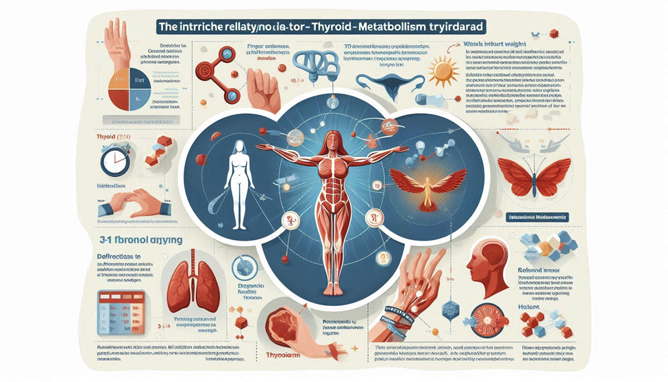
Iron is not merely a mineral used for hemoglobin; it is a structural and catalytic cofactor for dozens of metabolic enzymes, mitochondrial proteins, and most critically, for the synthesis of thyroid hormones. The thyroid gland, in turn, acts as the master regulator of basal metabolic rate (BMR), thermo genesis, lipid oxidation, carbohydrate turnover, protein synthesis, and mitochondrial respiration. When iron levels fall below optimal, thyroid hormone production, activation, and signaling become impaired. Mitochondria reduce ATP output, metabolic rate decreases, and the body naturally shifts toward energy conservation and fat storage.
This triad forms a metabolic loop:
Iron supports thyroid hormones → Thyroid hormones drive metabolism → Metabolism determines iron utilization and demand.
When any link in the triad weakens, the entire metabolic system experiences a slowdown. This is why individuals with iron deficiency—even without anemia—can experience:
- Unexplained weight gain
- Slower metabolic rate
- Fatigue and reduced physical activity
- Increased hunger and cravings
- Lower thermo genesis
- Impaired fat oxidation
- Poor exercise tolerance
- Disrupted glucose metabolism
Yet, millions of people with these symptoms never receive proper evaluation. Iron deficiency is one of the most common nutrient deficiencies worldwide, and thyroid disorders—especially subclinical hypothyroidism—is dramatically under diagnosed. When they coexist, they magnify each other’s metabolic consequences.
This guide explores the Iron–Thyroid–Metabolism Triad through a deeply scientific, mechanistic, dietitian-level lens, showing exactly how deficiency disrupts energy regulation, why it promotes weight gain, and how targeted nutrition and clinical strategies can restore metabolic balance.
1. Iron as the Metabolic Gatekeeper — Beyond Hemoglobin
Iron is one of the most metabolically influential micronutrients in the human body. While its role in oxygen transport is well known, far fewer people realize that iron is a required cofactor for:
- Thyroid peroxides (TPO) for thyroid hormone synthesis
- Deiodinase enzymes (DIO1, DIO2) for converting T4 → T3
- Cytochromes of the electron transport chain for ATP production
- Carnation synthesis for fatty acid transport
- Rib nucleotide reeducates for DNA replication and cellular turnover
- Catalane and peroxides for antioxidant defense
These functions make iron indispensable for metabolic rate, mitochondrial integrity, and endocrine regulation.
1.1 Iron as a cofactor in thyroid hormone production
The thyroid gland synthesizes two major hormones:
- T4 (thyroxin)
- T3 (triiodothyronine)
T3 is the active hormone that controls metabolic activity in nearly every tissue. The enzyme responsible for thyroid hormone synthesis—thyroid peroxides (TPO)—is iron-dependent. Without iron, TPO activity decreases, leading to:
- Reduced production of T4
- Reduced conversion of iodide to thyroid hormones
- Accumulation of unbound iodine
- Lower circulating thyroid hormone availability
This results in a physiologic state functionally identical to hypothyroidism, even if the thyroid gland itself is structurally healthy.
1.2 Iron and deiodinase function: The conversion problem
Even if the thyroid produces enough T4, the body still relies on selenium-dependent deiodinase enzymes to convert T4 → T3. These enzymes require iron-containing proteins and adequate iron stores to function efficiently.
Low iron reduces deiodinase activity, resulting in:
- High T4
- Low T3
- Normal TSH
- High reverse T3 (inactive)
This is the biochemical fingerprint often mistakenly labeled as “normal thyroid labs” despite clear signs of slowed metabolism. Weight gain in such cases is not due to caloric excess alone; it is a hormonal and mitochondrial consequence of impaired T3 signaling.
1.3 Iron and mitochondrial energy production
The electron transport chain (ETC) relies heavily on iron, which is embedded in cytochromes and iron–sulfur clusters. When iron is deficient, the ETC becomes inefficient.
Consequences include:
- Reduced ATP production
- Lower basal metabolic rate
- Reduced thermo genesis
- Impaired fatty acid oxidation
- Muscle fatigue and decreased activity levels
- Increased reactive oxygen species due to incomplete electron transfer
This leads to a scenario where the body produces less energy from the same amount of food, creating the sensation of fatigue, cold intolerance, and metabolic sluggishness.
2. Thyroid Regulation — the Hormonal Thermostat of Metabolism
Thyroid hormones regulate nearly every aspect of substrate metabolism:
- Resting metabolic rate
- Thermo genesis through brown fat
- Mitochondrial biogenesis
- Lipid utilization and biolysis
- Carbohydrate metabolism and insulin sensitivity
- Protein turnover
- Appetite and satiety signaling
Even mild reductions in thyroid hormone activity can decrease metabolic rate by 5–25%, depending on severity.
2.1 TSH is not enough: Why iron deficiency hides thyroid dysfunction
TSH is a pituitary hormone that responds to circulating levels of T4 and T3. But in iron deficiency:
- TSH may appear normal
- T4 may appear normal
- T3 is often low
- Reverse T3 is elevated
- Symptoms mimic hypothyroidism
This pattern is often dismissed because standard lab ranges fail to detect functional metabolic impairment. Iron deficiency masks hypothyroid patterns by limiting hormone production and conversion while keeping TSH artificially normal.
2.2 The body’s adaptive response: Energy conservation mode
When iron and thyroid hormones fall, the body shifts into energy conservation mode, characterized by:
- Lower BMR
- Reduced non-exercise thermo genesis (NEAT)
- Slower digestion
- Impaired fat mobilization
- Reduced muscle performance
- Increased fat storage
- Heightened hunger due to hormonal compensation
This mirrors the metabolic adaptation seen in chronic dieting, but with a biochemical root.
2.3 Thyroid hormones and brown adipose tissue (BAT)
T3 stimulates UCP1 in brown fat, increasing thermo genesis. Iron deficiency reduces T3 availability, causing:
- Reduced BAT activation
- Lower thermogenic output
- Increased cold sensitivity
- Lower calorie expenditure at rest
This is one of the clearest mechanisms linking iron deficiency to weight gain independent of caloric intake.
3. How Iron Deficiency Leads to Weight Gain the Mechanistic Chain
Iron deficiency affects weight through multiple converging pathways. It is not a single mechanism but a metabolic cascade, involving:
- Thyroid hormone suppression
- Mitochondrial dysfunction
- Hypoxia-induced metabolic adaptation
- Altered appetite regulation
- Reduced physical activity
- Poor glucose control
- Increased fat storage
- Reduced biolysis
- Impaired β-oxidation of fats
Below is a detailed breakdown of how these pathways interact.
3.1 Reduced thermo genesis and basal metabolic rate
Low iron → low T3 → reduced metabolic rate → fewer calories burned at rest.
Even a 10% reduction in BMR translates to ~150–300 fewer calories burned daily, equivalent to a weekly gain of 0.3–0.5 kg if intake remains unchanged.
3.2 Impaired fatty acid oxidation
Iron deficiency reduces:
- Carnation synthesis
- Cytochrome function
- β-oxidation enzymes
This forces the body to rely more on carbohydrates and store more fat. Even with a calorie-controlled diet, fat oxidation remains compromised.
3.3 Appetite deregulation
Iron deficiency increases:
- Gherkin (hunger hormone)
- Neuropeptide Y (NPY)
- Cortical
- Cravings for crabs and sugar
Simultaneously, lepton sensitivity decreases, making satiety signals weaker.
3.4 Fatigue → Reduced activity → lower energy expenditure
Iron-deficient individuals often report:
- Exercise intolerance
- Muscle pain
- Shortness of breath
- Weakness
This reduces structured exercise and spontaneous movement (NEAT), decreasing total daily energy expenditure by up to 300–600 calories.
4. Iron Deficiency without Anemia — the Hidden Metabolic Burden
Most people (including many clinicians) associate iron deficiency with anemia. However, iron deficiency progresses in three stages, and anemia is only the final stage. The earlier stages—known as Iron Deficiency without Anemia (IDWA)—are far more common and exert profound metabolic effects long before hemoglobin drops.
4.1 Stages of iron deficiency
- Stage 1 — Depleted iron stores (low ferreting)
- Hemoglobin normal
- Serum iron may fluctuate
- Thyroid and mitochondrial disruption already begin
- Stage 2 — Iron-deficient erythropoietin
- Transferring saturation drops
- TIBC rises
- Fatigue, cravings, cold intolerance appear
- Stage 3 — Iron-deficiency anemia (IDA)
- Hemoglobin falls
- Oxygen transport severely impaired
- Metabolic rate drops further
Most metabolic symptoms—including weight gain—occur in Stage 1 and Stage 2, because the body prioritizes hemoglobin production and diverts iron away from thyroid enzymes and mitochondria.
4.2 Why IDWA disrupts metabolism
In IDWA, the body still produces adequate hemoglobin, but enzymatic systems collapse first, particularly:
- TPO (thyroid hormone synthesis)
- Deiodinase enzymes (conversion to T3)
- Cytochrome proteins (ATP production)
- Carnation synthesis (fat transport)
This leads to:
- Lower T3
- Higher rT3
- Impaired mitochondrial function
- Reduced fat oxidation
- Decreased thermo genesis
Even without anemia, metabolic rate can drop by 10–20%, creating weight gain that appears “mysterious” or “unrelated to food intake.”
5. Why Women Are at Higher Risk — the Gender Metabolism Gap
Iron deficiency disproportionately affects women, with lifetime prevalence significantly higher than in men. This biological vulnerability amplifies the iron-thyroid interaction and increases the risk of metabolic slowdown.
5.1 Menstruation and iron losses
Monthly blood loss removes iron directly from circulation. Heavy menstrual bleeding (HMB), even when considered “normal,” can cause:
- Chronic ferreting depletion
- Lower T3 production
- Increased fatigue
- Disrupted metabolic rate
- Poor lipid metabolism
- Increased desire for high-crab foods during the lacteal phase
Women with ferreting below 40–50 nag/mol often report weight gain even with unchanged diet and exercise patterns.
5.2 Pregnancy and postpartum demands
Pregnancy increases iron requirements dramatically because iron is needed for:
- Fetal brain and thyroid development
- Maternal blood volume expansion
- Placental growth
- Mitochondrial biogenesis
Even mild iron deficiency increases:
- Gestational fatigue
- Insulin resistance
- Fatigue-driven lower activity
- Higher likelihood of postpartum weight retention
Postpartum iron depletion also contributes to:
- Hair loss
- Cold intolerance
- Low metabolic rate
- “Baby weight” that is resistant to dieting
5.3 Thyroid autoimmunity (Hashimoto’s) and iron status in women
Women are 8–10× more likely to develop autoimmune thyroid disease. Iron deficiency plays a role by:
- Impairing TPO, making thyroid tissue more vulnerable
- Increasing oxidative stress in the gland
- Altering immune tolerance
- Stressing thyroid hormone synthesis
Women who have both Hashimoto’s and low iron often struggle with:
- Weight gain
- Intolerance to exercise
- Constipation
- Chronic fatigue
- Difficulty losing weight despite strict diets
This combination is frequently overlooked, leading to misdiagnosis or under-treatment.
6. Inflammation, Hepcidin, and Metabolic Blockade
One of the most overlooked aspects of the Iron–Thyroid–Metabolism Triad is inflammation, which regulates iron transport through a hormone called hepcidin.
6.1 Hepcidin: The master controller of iron absorption
Hepcidin is produced by the liver and controls iron availability by:
- Blocking intestinal absorption
- Trapping iron inside cells (especially macrophages)
- Preventing iron release into the bloodstream
High hepcidin = low available iron, even if dietary intake is high.
6.2 What increases hepcidin?
- Chronic stress
- Inflammation
- Obesity
- High cortical
- High insulin
- Illness
- Poor sleep
- Excessive exercise without recovery
- High sugar diets
This creates a metabolic paradox:
People can consume iron-rich foods but still be deficient because inflammation blocks absorption.
6.3 Obesity and hepcidin
Adipose tissue produces inflammatory cytokines (IL-6, TNF-α) that raise hepcidin levels.
This creates a vicious cycle:
Obesity → Inflammation → High hepcidin → Low iron → Lower thyroid → slower metabolism → more fat storage.
6.4 Hypothyroidism and inflammation: A feedback loop
Low thyroid hormone increases:
- Gut permeability
- Inflammatory cytokines
- Lipid accumulation in tissues
Inflammation increases hepcidin, which blocks iron absorption, suppresses TPO, and worsens thyroid dysfunction. This feedback loop causes progressive metabolic suppression.
7. Diagnostic Markers — Why Standard Blood Tests Miss the Problem
Most patients with metabolic symptoms receive only a basic panel:
- Hemoglobin
- Hematocrit
- TSH
These tests are insufficient for diagnosing metabolic iron–thyroid issues.
7.1 Comprehensive iron panel required
A true metabolic evaluation requires:
- Ferritin
- Serum iron
- Transferrin saturation (TSAT)
- TIBC (Total Iron Binding Capacity)
- star (soluble transferring receptor)
- CRP (inflammation marker)
- Hepcidin (optional but useful)
7.2 Functional ferreting ranges
Although labs often consider ferreting of 10–20 nag/mol “normal,” metabolic studies show:
- < 30 nag/mol — iron deficiency
- 30–70 nag/mol — suboptimal for thyroid
- 70–120 nag/mol — ideal for thyroid conversion and metabolism
- 150 — often inflammation-driven iron trapping
Many individuals stuck at ferreting 12–30 experience:
- Fatigue
- Weight gain
- Low body temperature
- Poor exercise tolerance
- Hair thinning
- Sleep disruption
7.3 Thyroid panel needed beyond TSH
A metabolic thyroid evaluation includes:
- TSH
- Free T4
- Free T3
- Reverse T3
- TPO & TG antibodies
- SHBG (Thyroid-dependent marker)
7.4 The iron–thyroid signature pattern
Many people have this metabolic pattern:
- Normal TSH
- Low or low-normal Free T3
- High reverse T3
- Ferreting < 70
- Low TSAT
- Normal hemoglobin
- Fatigue + weight gain
This is functional hypothyroidism caused by iron deficiency, not a psychiatric or “lifestyle compliance” issue.
8. Diet, Absorption, and the Metabolic Iron Recovery Plan
Iron intake matters — but absorption is the real bottleneck. Only 10–15% of dietary iron is absorbed under ideal conditions.
8.1 Two types of iron
Hemet iron
- Found in meat, poultry, fish
- Absorption: ~20–35%
- Little affected by inhibitors
Non-home iron
- Found in plants
- Absorption: 1–10%
- Strongly impacted by enhancers and blockers
8.2 Foods that enhance absorption
- Vitamin C (citrus, kiwi, berries, peppers)
- Animal proteins
- Fermented foods (lower phytates)
- MFP factor from meat/fish
- Acidic foods (lemon juice, vinegar)
8.3 Foods that inhibit absorption
- Coffee and tea (polyphones)
- Calcium supplements
- High-fiber raw grains
- Phytates (in beans, legumes, whole grains)
- Oxalates (spinach, almonds)
- Certain medications (PPIs, antacids)
Timing matters:
Avoid inhibitors for 1–2 hours around iron-rich meals.
8.4 Iron-rich foods for thyroid and metabolism
Hemet sources (best absorbed):
- Beef
- Lamb
- Chicken liver
- Turkey
- Sardines
- Tuna
- Oysters
- Mussels
Non-home sources (needs vitamin C):
- Lentils
- Beans
- Pumpkin seeds
- Tofu
- Spinach (limited absorption)
- Quinoa
8.5 The ferreting recovery timeline
- 4–6 weeks: energy improves
- 8–12 weeks: metabolic rate increases
- 3–6 months: ferreting reaches optimal range
- 6–12 months: full thyroid–metabolic recovery
Conclusion
Iron, thyroid function, and metabolic regulation form a tightly interconnected physiologic network that determines how efficiently the body produces energy, regulates body temperature, maintains metabolic flexibility, and manages body weight. When iron availability is compromised—whether through inadequate intake, poor absorption, and chronic inflammation, or increased physiological demand— thyroid hormone synthesis becomes impaired. This reduction in active thyroid hormones disrupts mitochondrial ATP production, lowers basal metabolic rate, and alters fuel utilization patterns, creating a metabolic environment that favors fatigue, reduced thermo genesis, impaired fat oxidation, and gradual weight gain.
The challenge is compounded by the fact that early iron deficiency and subclinical hypothyroidism often present with nonspecific symptoms—fatigue, cold intolerance, hair changes, and cognitive fog— leading many cases to remain undiagnosed for years. For individuals struggling with unexplained weight fluctuations, stubborn weight loss resistance, or chronic low energy, investigating iron status (serum ferreting, iron saturation, transferring receptor) and thyroid markers (TSH, free T4, free T3, reverse T3, antibodies) can uncover hidden metabolic blocks that standard assessments often miss.
From a therapeutic perspective, restoring iron sufficiency is not merely about supplementation—it requires personalized dietary strategies, improved absorption practices, inflammation reduction, gut health optimization, and addressing menstrual or lifestyle factors that perpetuate iron loss. Likewise, supporting thyroid function involves aligning nutrient intake (iodine, selenium, and zinc), circadian rhythms, stress regulation, and metabolic timing to ensure hormonal stability.
Ultimately, the iron–thyroid–metabolism triad underscores a central truth: metabolic health is multifactorial, and weight regulation extends far beyond calories. Recognizing this triad allows clinicians, dietitians, and individuals to adopt targeted, biologically informed interventions that correct root causes, restore metabolic efficiency, and support sustainable long-term weight management.
SOURCES
World Health Organization, 2024. Micronutrient Deficiencies: Iron Deficiency and Anemia.
Beard & Connor, 2023. Iron status and thyroid hormone metabolism: A biochemical review.
Zimmermann & Boehlert, 2020. Iodine and thyroid hormone synthesis physiology.
Camaschella, 2019. Iron deficiency: Molecular mechanisms and diagnostics.
Botanic et al., 2022. Subclinical hypothyroidism and metabolic outcomes.
Lazarus, 2021. Thyroid function, energy expenditure, and weight regulation.
Allen, 2020. Iron bioavailability and dietary absorption: Clinical implications.
Gang, 2020. Hepcidin regulation and inflammatory control of iron absorption.
Mancini et al., 2023. Thyroid hormones and mitochondrial bioenergetics.
Soma & Pirate, 2021. Iron-deficiency anemia and chronic fatigue physiology.
Varna et al., 2019. Iron loss mechanisms in women: A clinical overview.
Galton, 2022. T4-to-T3 conversion: Nutritional and metabolic determinants.
Hider & Silliman, 2019. Iron deficiency and its systemic metabolic effects.
O’Leary & Sampan, 2020. Iron interactions with zinc, calcium, and phytates in diets.
Juntas & Jonklaas, 2022. Diagnosis and management of hypothyroidism.
Reddy & Sod, 2021. Iron deficiency and weight-gain tendencies: Review of mechanisms.
Zimmermann, 2018. Micronutrients and thyroid gland physiology.
Lopez et al., 2023. Iron supplementation strategies: Efficacy and tolerability.
Cheng & Canaries, 2020. Thyroid disease prevalence and metabolic consequences.
Petri et al., 2021. Global patterns of iron deficiency across populations.
Morris & Johnston, 2022. Gut health and iron absorption: Clinical correlations.
Harvey & Williams, 2019. Thyroid hormones and resting metabolic rate regulation.
Hatcheck & Shoo, 2019. Safety evaluation of iron and micronutrient supplementation.
Rochester et al., 2023. Low ferreting and metabolic adaptation in women of reproductive age.
Seth & Raj put, 2022. Nutrient–endocrine interactions in thyroid disorders.
HISTORY
Current Version
Nov 24, 2025
Written By
ASIFA
0 Comments